The acromioclavicular (AC) joint is located at the apex of the shoulder where the portion of the scapula called the acromion articulates with the clavicle. It has a cartilaginous structure with a shock-absorbing function called the meniscus; its stability depends on the coraco-clavicular (CC) ligaments (Fig. 1).

The AC joint is generally injured following falls resulting in direct trauma to the shoulder: the scapula is pushed down while the clavicle tends to remain in position. [1]

The loss of contact between the bone heads is termed acromioclavicular (AC) dislocation and there are 6 different types based on severity according to Rockwood’s classification [2]. (Fig.2)

Riparazione di lussazione acromion-claveare
Fig. 1 Normal shoulder without ligament lesions: detail of the acromioclavicular (AC) joint.
Fig. 2 Acromioclavicular (AC) dislocation with rupture of the AC joint capsule and injury to the coraco-clavicular (CC) ligaments.

The symptomatology is characterised by pain associated with partial or total functional inability of the limb.

The diagnosis is made following a specialist clinical examination (Fig. 3), which also makes use of instrumental examinations such as X-rays (Fig. 4), ultrasound and nuclear magnetic resonance imaging (MRI).

Fig. 3 Clinical appearance of a patient after AC dislocation. One can see the dysmetria of the left shoulder in relation to the right with the typical 'piano key' clavicle sign.
Fig. 4 Radiographic evaluation of a patient after AC dislocation. The loss of contact of the clavicle with the acromion of the scapula can be seen.

Treatment may be conservative or surgical.

conservative treatment is reserved for grade 1 and 2 dislocations. Grade 3 injuries, on the other hand, can be treated conservatively by assessing factors such as occupation, dominant hand, type of work or sport, risk of re-injury, etc. Conservative therapy involves rest, anti-inflammatory and pain-relieving drugs, and appropriate physiotherapy.

Surgical treatment is reserved for grade 3 dislocations that are non-responsive/unsuitable for conservative therapy, grade 4,5 and 6 dislocations [3,4].

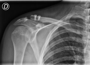
The technique we use for the reconstruction of this joint makes use of very strong artificial ligaments (LARS) used in a minimally invasive technique to reconstruct the DC ligaments, thereby stabilising the acromioclavicular joint. (Fig.5 and 6)

Fig. 5 Minimally invasive AC dislocation stabilisation surgery using an artificial ligament (LARS) to reconstruct the DC ligaments.
Riparazione di lussazione acromion-claveare
Fig. 6 Post-operative X-ray check after AC stabilisation by minimally invasive technique.


The surgery is followed by a specific rehabilitation physiotherapy protocol. The results are very good with a success rate of 98%. [5]

Bibliography
  1. The Diagnosis and Treatment of Acute Dislocation of the Acromioclavicular Joint. Frank Martetschläger, et al. Dtsch Arztebl Int. 2019 Feb 8;116(6):89-95.
  2. Fracture classification systems: do they work and are they useful? Rockwood CA Jr. J Bone Joint Surg Am. 1994 May;76(5):790.
  3. Rockwood type III acromioclavicular dislocation: surgical versus conservative treatment. Gstettner C, Tauber M, Hitzl W, Resch H.
    Shoulder Elbow Surg. 2008 Mar-Apr;17(2):220-5.
  4. Return to sport after surgical treatment for high-grade (Rockwood III-VI) acromioclavicular dislocation. Verstift DE, Welsink CL, Spaans AJ, van den Bekerom MPJ. Knee Surg Sports Traumatol Arthrosc. 2019 Dec;27(12):3803-3812.
  5. Acromioclavicular joint reconstruction with the LARS ligament in professional versus non-professional athletes. Marcheggiani Muccioli GM, Manning C, Wright P, Grassi A, Zaffagnini S, Funk L. 2016 Jun;24(6):1961-7.

Book online